Nurturing developing neurons: How microglia, the main immune cells of the brain contribute to brain development by interacting with newborn neurons
The “Lendület” – Momentum Laboratory of Neuroimmunology of the ELKH Institute of Experimental Medicine (IEM) led by Ádám Dénes was the first to describe the presence of a direct link between microglia cells and the cell body of developing neurons. The researchers also revealed the role of these contacts in brain development. The discovery may be of crucial importance for understanding the mechanisms of brain immune processes that often underlie neurodevelopmental disorders and related neuropathologies. The results of the study have been published in the prestigious international journal Cell Reports.
Microglia are recognized by the scientific community as the main immune cells of the central nervous system and the main regulators of inflammatory processes in the brain. The involvement and altered function of microglia have been demonstrated in the pathogenesis of most neurological diseases, such as stroke, Alzheimer’s disease, Parkinson’s disease, epilepsy, dementia and amyotrophic lateral sclerosis (ALS). The role of inflammatory processes and microglia in neurodevelopmental disorders is also increasingly recognized. The research group has accumulated a substantial body of knowledge in the field of microglia-neuron interactions, with several internationally visible publications in recent years. Among others, the group has discovered a novel form of interaction between microglia and the cell body of neurons, named somatic junctions, and revealed their role in microglia-mediated neuroprotection in the adult brain. Although the important role of the microglia in brain development has been pointed out in many previous studies, it was not known, which cell-to-cell communication mechanisms allow microglia to influence neuronal development and the formation of complex neuronal networks.

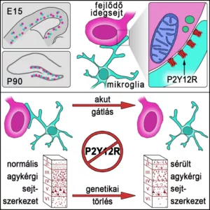
Microglial processes establish direct contacts on the cell bodies of developing neurons from embryonal day 15 (E15), throughout postnatal development and during postnatal neurogenesis. These contacts possess a specialized structural and molecular composition, as neuronal mitochondria and vesicles are enriched at these sites, with the accumulation of the microglia-specific P2Y12-receptors on contacting microglial processes. Acute inhibition of these receptors leads to inhibition of contact formation, while genetic deletion of P2Y12 receptors results in disturbed cortical cytoarchitecture.
Among the members of Ádám Dénes’ research group, Csaba Cserép and his student Dóra Anett Schwarcz played an outstanding role in the implementation of the research program, with further contribution by the research group of István Katona at IEM. During their investigations, the researchers used both high-resolution molecular anatomy techniques, combined light- and electron microscopy, and ex vivo imaging studies. Using a multifaceted approach, the researchers proved the presence of direct connections between microglia and developing neurons both during embryonic development and after birth. The specific, dynamically changing anatomical connections between microglia and developing, immature neurons resemble in many respects somatic junctions previously discovered by the research group, and their unique structure allows microglia to continuously monitor and effectively influence the development and networking of neurons. The significance of this discovery is illustrated by the fact that blockade of communication through these interactions sites prevents the development of normal cortical structure. Microglia should therefore be regarded as an important regulatory cell type in brain development, and a better understanding of their function may help in the development of therapies for neurodevelopmental disorders and other conditions.

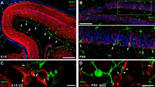
A) Confocal laser scanning microscopic image of embryonal brain shows the distribution of microglia (green) and developing neurons (red). White arrows point to the enrichment of microglia. B) Microglia (green) are enriched in the subgranular zone of the hippocampal dentate gyrus. Developing neurons (red) are contacted by microglial processes, mature granule cells are blue. C-D) High resolution images show some examples of microglia-contacts in embryonal (C) and the adult brain (D), white arrows point to contact sites. Scale bar is 200 µm on A, 100 µm on B, 5 µm on C and 10 µm on D.
This research was supported by the following grants:
ERC-CoG 724994, the Hungarian Academy of Sciences “Lendület” – Momentum Programme LP2016-4/2016 and LP2022-5/2022, the Hungarian-Chinese Applied Research and Development Cooperation Programme 2020-1.2.4-TÉT-IPARI-2021-00005, the Hungarian Academy of Sciences NKFIH. Csaba Cserép (UNKP-20-5) and Balázs Pósfai (UNKP-20-3-II) have also received funding from the National R&D Funding Programme of the Hungarian National Research Council. Anett Scwarcz received a grant from the Richter Gedeon Talentum Foundation. István Katona was supported by the National Brain Research Programme (2017-1.2.1-NKP-2017-00002) and the National Research and Technology Foundation’s Lifeline Programme (129961).
Publication:
Csaba Cserép*, Anett D. Schwarcz, Balázs Pósfai, Zsófia I. László, Anna Kellermayer, Zsuzsanna Környei, Máté Kisfali, Miklós Nyerges, Zsolt Lele, István Katona, Ádám Dénes* (2022). Microglial control of neuronal development via somatic purinergic junctions. Cell Reports. doi: 10.1016/j.celrep.2022.111369

